


Description of HE project
DESCRIPTION OF THE RESEARCH PROJECT J3-1748 (Role of MRI in detection of minimal hepatic encephalopathy)
Background:Most common cause of liver cirrhosis (LC) is alcohol overconsumptionand its incidence increases. One of the complications of the cirrhosis ishepatic encephalopathy (HE), defined as brain dysfunction caused byliver insufficiency. Since HE is potentially reversible, the recognition ofearly stages is crucial. Minimal HE (mHE) is defined as HE withoutsymptoms on clinical examination, but can be detected bypsychometric testing. Early recognition of mHE is pivotal as earlytreatment improves the quality of life of patients.Pathophysiological mechanisms of HE are complex. Ammonia,cytokines, manganese deposition and neurotransmittersare involvedin malfunctioning of neuronal cells, but exact mechanisms are poorlyunderstood.
2. Problem and objectives of the proposed researchThis study will address the following problems:Diagnosis of mHEDiagnosis of mHE is challenging, time-consuming and subjective. MRIand MRS are non-invasive and objective methods that producequantifiable results. This project will assess the role of MRI in mHEdiagnosis with emphasis on multimodal imaging technique. Thecorrelation of psychometric testing with MRI data will improve thesensitivity and the specificity of mHE diagnosis.Pathophysiology of HEThe exact mechanisms of neuron and astrocyte damage in HEarepoorly understood. Advanced MRI and MRS techniques will providenew answers to these questions:
2.1. Detection of minimal brain water shifts, paramagnetic deposits inmHE and HE involved in pathophysiological mechanisms of HEdevelopment.With advanced MR techniques such as DKI and QSM,in-vivo detections of small water shifts content, intracerebraledemaand brain deposits are feasible. This will also offer the possibility tocompare and evaluate the results from previous studies on animalmodels and cell cultures
2.2. Assessment of the role of GABA in mHE and HE. Striatum has acrucial role in movement planning, decision-making, motivation andreward processes With MEGA-PRESS, millimolar concentrationof GABA,the most important inhibitory neurotransmitter in the brain, can bereliably detected. This will provide evidence for early and lateimpairment of striatum in mHE/HE.2.3. The potential use of various MR techniques in detecting mHE inclinical environment.MR is a non-invasive and safe method thatcould have an essential role in detecting mHE. We will assess thesensitivity and specificity of MR imaging in mHE diagnosis withemphasis on multimodal imaging technique.
3. The originality of the expected results
Multimodal MRI approach as we propose in this application has notbeen yet performed. We will use advanced MRI techniques which arecurrently not available in clinical setting. QSM requires multicentercollaboration.Partners at MUG will assess the magnetic properties of white matterlesions and normal appearing white matter (NAWM) in patients withhepatic encephalopathy (HE) and minimal HE (mHE). Alterations ofthe magnetic susceptibility will be compared to control tissue andrelated to clinical status. Quantitative susceptibility maps (QSM) willbe calculated
with a home developed total generalized variation (TGV)approach, which incorporates individual steps of phase unwrapping,background field removal and dipole inversion in a single iteration andtherefore yields a robust solution to the reconstruction problem. TheTGV algorithm was developed for Siemens data and therefore willbeadapted to process also raw data from the Philips scanner in Ljubljanaduring the first year of the project. In addition to QSM, we will alsoassess R2* in lesions and NAWM. R2* will be calculated from multiechodata and corrected for dephasing effectsfrom background fields.Adaption and optimization of the QSM and R2* calculation pipelinesand the image analysis of the corresponding maps will be done by aPh.D. student with half effort in two years.
4. Methods
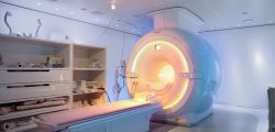
Patients with hyperammonemia and mHE and patients with HE will bestudied. Diagnosis of HE will be based on results of validatedneuropsychiatric test listed below. Age-matched and gender-matchedvolunteers will serve as the control group.Patients with contraindications for MRI scanning will beexcluded. Thestudy protocol will include: 1) short description and the aim of thestudy and patient’s written-consent, 2) scoring according to W H and CPclassifications, 3) liver function tests, ammonia levels, 4) CriticalFlicker Frequency test, 5) neuropsychiatric tests and 6) MRIscanning.Standard safety questionnaire will be used before the scanning. MRIscanning will involve: T1-and T2-weighted MRI, MRS (MEGA-PRESSand PRESS), high resolution diffusion weighted imaging (DW I),diffusion kurtosis imaging (DKI), quantitative susceptibility mapping(QSM). Liver QSM will be executed to assess iron load. The sametests will be performed on control subjects. The advanced MRI, withemphasis on complex analysis, will be done with collaboration ofNeuroimaging Research Unit, Medical University Graz.
6. Relevance and potential impact of the results
This project will explore mechanisms that are complex and still poorlyunderstand in humans. Multimodal MR approach that will be used inprotocol is novel approach to study HE. Moreover, as the diagnosis ofmHE is time-consuming and often delayed or even unrecognized, MRcould be used as method in a process of diagnostic process of mHE.
7. Organizational structure and feasibility of the project
Acquisitionof MRI and MRS will be performed at ULMF, analysis ofQSM results will be done in collaboration with MedUni Graz andoptimization of protocols and analysis of structural data jointly withAcademic Research Centre of SZD.
8. Comments
Collaboration with Austrian partner is essential for QSM data analysis